
In the 150 years since chromosomes were first discovered, scientists have studied them in various contexts, from insect cells to artificial systems.
Chromosomes, first discovered in 1882, are involved in various processes from genome organization to cancer and autoimmunity.
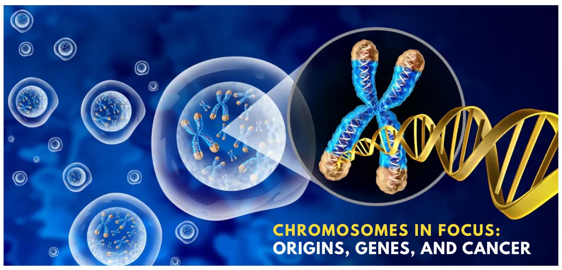
Human DNA, which is approximately two meters in length, is organized into a compact, thread-like structure known as the chromatin, so it could fit into the nucleus, a tiny sphere that’s only about six millionths of a meter in diameter. Before cells divide, the chromatin is condensed even further into chromosomes. These structures, first discovered by the German biologist Walther Flemming in 1882, are visible as dark blobs under the microscope during cell division. Human cells have 22 pairs of “autosomal” chromosomes and a pair of sex chromosomes. Learn how these chromosomes were named and catch up on the latest chromosome stories here.
The Origins of X and Y Chromosome Nomenclature
Calling the sex chromosomes “X” and “Y” may seem unfair to chromosome numbers 1 to 22. But the designation of sex chromosomes using letters, rather than numbers, dates all the way back to the late 1800s. In 1891, the German biologist Hermann Henking saw a large chromosome in the firebug sperm cell and called it “X element”, though at the time, scientists had not yet known that specific chromosomes could determine the sex of organisms. In the decades that followed, other researchers who studied sex cells in other animals, from beetles to mammals, continued this tradition. Then, in 1960, an international panel of researchers convened to decide on a naming system for human chromosomes—they agreed to keep the “superfluous appellation” of the sex chromosomes, and the rest was history.
Plastic Additive Linked to Chromosome Abnormality
When hermaphrodite organisms like the roundworm Caenorhabditis elegans self-fertilize, males only arise due to a cell division error during the formation of sex cells. This phenomenon, called X chromosome non-disjunction, produces cells with abnormal numbers of X chromosomes, which in humans, could result in genetic disorders such as the Turner and Klinefelter syndromes. In 2024, Monica Colaiácovo, a molecular and cell biologist at Harvard Medical School, exposed C. elegans to benzyl butyl phthalate (BBP), a common plastic additive, at concentrations comparable to what have been previously reported in pregnant women. Colaiácovo found that in response to this treatment, the proportion of male C. elegans rose from 0.2 percent to a whopping 30 to 40 percent, indicating a dramatic increase in the incidence of X chromosome non-disjunction.
Sex Chromosome Loss Linked to Cancer Susceptibility
During early female development in humans, one of the two X chromosomes gets inactivated so that XX females and XY males end up with a similar number of X chromosome gene products. As genetically female individuals age, they could undergo further X chromosome inactivation. This process, known as mosaic loss of X (mLOX), can cause blood cells to divide abnormally and increase susceptibility to various diseases, including cancer. Recently, a study that involved 900,000 genetically female individuals reported that up to 12 percent experienced mLOX, and this phenomenon is indeed associated with cancer susceptibility. The researchers, led by Mitchell Machiela, a genetic epidemiologist at the National Cancer Institute, also found that while Y chromosome inactivation in XY males is similarly associated with blood cell abnormality and cancer, the loss of X and Y chromosomes later in life likely occur through different molecular mechanisms.
X Chromosome Inactivation Is a Double-Edged Sword
X chromosome inactivation is an important part of XX female development. However, about a quarter of the genes that are meant to be silenced can “escape” this process, and scientists have found that this evasion can be a double-edged sword. In some cases, escape from X chromosome inactivation can protect genetically female individuals from cancer, while in others, it can increase their risk for developing autoimmune disorders such as multiple sclerosis and lupus.
X Chromosome Inactivation Product Promotes Autoimmunity
Researchers have observed that people with Klinefelter syndrome, who have a set of active XY chromosomes and an extra, inactive X chromosome, are more prone to female-biased autoimmune disorders, such as lupus. In a 2015 study, Howard Chang, a dermatologist at Stanford University, identified 81 proteins that bind to X-inactive specific transcript (XIST), the long noncoding RNA that results from X chromosome inactivation. More recently, Chang noticed that some of these proteins overlapped with those on the list that physicians use to diagnose autoimmune disorders. He hypothesized that the interaction between the autoimmune-associated proteins and XIST underlie female-biased autoimmunity. Then, in a 2024 study, now published in Cell, Chang and his colleagues showed that male mice that were genetically engineered to express XIST produced more autoantibodies.
Chaotic Chromosomes Drive Cancer Progression
Unlike normal human cells, which have two copies of each chromosome, cancer cells can have up to five or six as a result of the genomic chaos commonly known as chromosome instability (CIN). The role of CIN in cancer was first described in 1997, but for a long time, it was overshadowed by research efforts that focused on specific genetic mutations, thanks to the rise of new sequencing methods. In 2010, scientists at the Memorial Sloan Kettering Cancer Center, led by Robert Benezra, showed that CIN could eliminate cancer’s dependency on the oncogene caused it in the first place. This study revived scientists’ interest in CIN, and since then, researchers have uncovered various molecular mechanisms that underlie this relationship.
Chromosome Lagging Strand Has Its Own “Telomerase”
The end-replication problem refers to DNA polymerase’s inability to replicate the ends of linear chromosomes. To prevent the loss of valuable genetic information due to chromosomes shortening with every cell division, cells have two well-known protective mechanisms: telomeres, regions of repetitive sequences at the ends of chromosomes, often likened to the protective caps that prevent shoelaces from fraying, and telomerase, the enzyme that replenishes telomeres by adding single-stranded nucleotide repeats to telomere ends. Recently, Titia de Lange, a cell biologist and geneticist at Rockefeller University, and her colleagues found that while telomerase solves the end-replication problem in the leading strand, the lagging strand relies on a different solution. In their 2024 study, published in Nature, de Lange’s team found that the lagging strands in cells that lack the CST-Polα-primase protein complex were unable to keep up with DNA replication, indicating this complex’s role as the lagging strand’s “telomerase.”
Artificial Chromosome Synthesis Got a Bigger Start
Human artificial chromosomes (HACs) can deliver engineered DNA into cells and behave like natural chromosomes, offering a promising approach for both research and therapeutic purposes. HACs are especially useful in circumstances that require the replacement of a large gene. To synthesize HACs, scientists typically link shorter DNA fragments together. However, this approach is limited because the short fragments often come together unpredictably. Recently, researchers at the University of Pennsylvania, led by biochemist Ben Black, overcame this issue by starting with DNA fragments that were more than three times the size of the ones typically used.